
Groin Injury
Definition
Pain in the groin: the junction of the thigh and the abdomen.
Classified using the Doha Agreement (2015): a clinical taxonomy based on history and examination, not imaging.
Epidemiology
- 4 to 19% of all time-loss injuries in male football
- Roughly 4 players in a squad of 25 elite footballers sustain a time-loss groin injury each season
- Highest risk in sports with kicking, sprinting and rapid change of direction: football, rugby, Australian football, ice hockey, hockey
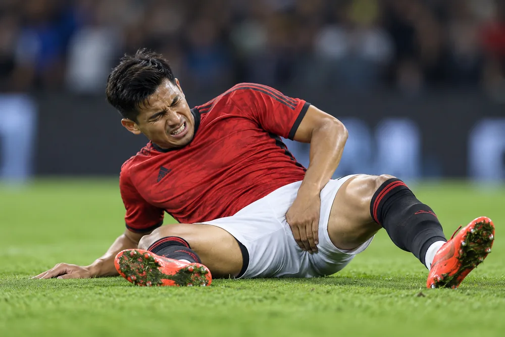
Relevant Anatomy
The pubic symphysis is the central anchor. Forces from the lower limb and trunk converge here.
- Adductors: adductor longus, brevis, magnus, pectineus, gracilis. Adductor longus originates from the anterior pubic body, just below the pubic tubercle
- Hip flexors
- Iliopsoas: psoas (T12 to L4 vertebrae) and iliacus (iliac fossa) to the lesser trochanter
- Rectus femoris: AIIS and superior acetabulum to patella and tibial tuberosity (via patellar tendon)
- Sartorius: ASIS to proximal medial tibia
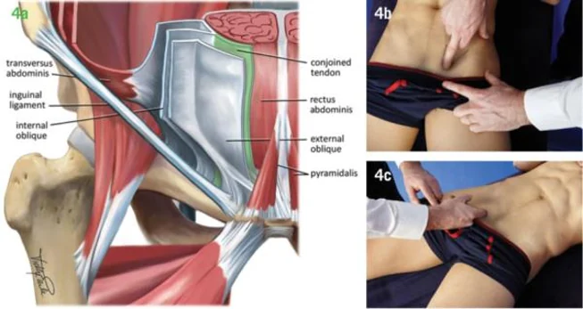
- Inguinal canal: superficial ring, conjoint tendon, posterior wall (transversalis fascia)
- Rectus abdominis: inserts on the pubic crest, sharing an aponeurosis with adductor longus
- Hip joint: deep groin pain source
Pathophysiology
- Repetitive high-load activities: kicking, sprinting, change of direction, stretching into end range
- Imbalance between adductor and abdominal forces across the pubic symphysis
- Progressive overload of tendon, muscle, bone or the inguinal wall
Risk Factors
| Intrinsic | Extrinsic |
|---|---|
| Previous groin injury (strongest predictor) | Type of sport (kicking, change of direction) |
| Reduced hip adductor strength | High training and match load |
| Reduced hip range of motion | Higher level of play |
| Previous lower limb injury | Pre-season and congested match schedules |
Classification (Doha Agreement 2015)
1. Defined clinical entities
Doha is the most widely used in sports medicine.
| Entity | Key features | Diagnostic tests |
|---|---|---|
| Adductor-related | Most common. Adductor longus most affected | Adductor tenderness + pain on resisted adduction |
| Iliopsoas-related | Second most common in many series. Iliopsoas, rectus femoris, sartorius | Iliopsoas tenderness: more likely if pain on resisted hip flexion and/or hip flexor stretch |
| Inguinal-related | No palpable inguinal hernia | Pain in inguinal canal region + tenderness: more likely if pain on resisted abdominal testing, Valsalva, cough or sneeze |
| Pubic-related | Pubic bone stress | Local tenderness of the pubic symphysis and adjacent bone. No specific resistance test |
2. Hip-related groin pain
- FAI syndrome, labral tears, chondral injury, hip OA, AVN.
3. Other causes of groin pain
- Inguinal or femoral hernia
- Nerve entrapment (obturator, ilioinguinal, genitofemoral)
- Referred pain: lumbar spine, sacroiliac joint
- Intra-abdominal and urological pathology
- Scrotal pathology: testicular torsion, epididymitis, varicocele
Other consensus frameworks exist:
- Manchester 2014 (British Hernia Society, “inguinal disruption”)
- Milan Agreement 2016 (“groin pain syndrome”)
Clinical Presentation and Assessment
History
- Location guides diagnosis
- Medial groin, radiating down inner thigh: adductor-related
- Anterior thigh / anterior groin: hip flexor-related
- Lower abdomen, inguinal canal: inguinal-related
- Central, over the symphysis: pubic-related
- Deep groin: hip joint
- Buttock: lumbar spine, SIJ, deep gluteal syndrome
- Mechanism: kicking, change of direction, sprinting, stretching
- Onset: acute (strain, avulsion) or gradual (overuse)
- Snapping or “pop” at injury: consider avulsion or complete tear
- Training history and load changes
- Red flags: fever, night pain, weight loss, urinary or scrotal symptoms
Examination
Principle: Press, Stretch, Resist for each structure.
- Palpation of each anatomical landmark
- Stretch tests: passive adduction stretch; modified Thomas test for hip flexors
- Resistance tests: squeeze tests, resisted hip flexion, resisted sit-up
- Hip joint exam: range of motion, FADIR, FABER
- Hernia examination standing and supine, including cough impulse
Investigations
- Pelvic radiograph: avulsion fractures, FAI morphology, pubic symphysis changes, OA
- Ultrasound: tendon and muscle injury, dynamic hernia assessment, guided injections
- MRI: muscle-tendon injury, pubic bone marrow oedema, stress fracture, hip pathology
- Imaging findings are common in asymptomatic athletes. Interpret against clinical findings
1) Adductor-Related Groin Pain
- Most common entity
- Adductor longus most often affected, at the proximal musculotendinous junction or the enthesis on the pubic bone
- About a third bilateral
Clinical features
- Medial groin pain at the adductor longus origin, radiating down the inner thigh
- Pain with sprinting, sharp change of direction and kicking
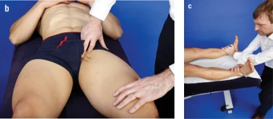
- Palpate adductors with the hip in abduction and external rotation (frog position)
- Squeeze tests
- Long-lever: legs extended, examiner’s hand between the ankles
- Short-lever: hips 45° flexion, fist or ball between the knees
- Pain and/or reduced force are positive
- Pain on passive adductor stretch
Treatment
- Relative rest; avoid pain-provoking activity
- Progressive strengthening: isometric, then concentric, then eccentric
- General dynamic stabilisation of pelvic and core muscles
- Hölmich active training programme (exercise-based, 8 to 12 weeks)
- Module I (weeks 0 to 2): static adduction against a ball, sit-ups, combined abdominal and hip flexion, balance board work
- Module II (week 3 onwards): side-lying abduction/adduction, back extensions, standing one-leg weighted pulley adduction/abduction, coordination and sliding board exercises
- Return to play: around 6 to 12 weeks for longstanding cases
2) Iliopsoas-Related (Hip Flexor-Related) Groin Pain
Mechanism
- Kicking and sprinting: rectus femoris
- Change of direction: iliopsoas
Clinical features
- Anterior groin and anterior proximal thigh pain
- Pain after deep sitting or climbing stairs
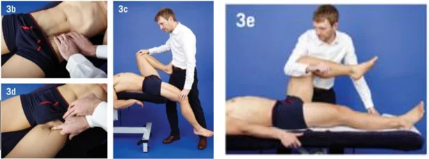
- Palpation
- Iliopsoas: just medial to the ASIS and below the inguinal ligament, palpated deeply during full expiration
- Rectus femoris: from the AIIS distally
- Resisted hip flexion
- Iliopsoas: hip at 90° flexion
- Rectus femoris: knee extended
- Pain on hip flexor stretch: modified Thomas test
Treatment
- Same general principles as adductor-related pain
- Specific hip flexor strengthening, including eccentric iliopsoas work in the inner range
- Return to play: around 3 to 6 weeks
3) Inguinal-Related Groin Pain
Also termed inguinal disruption, sportsman’s groin, sports hernia, athletic pubalgia. Thought to involve weakness of the posterior inguinal wall, with bulging during raised intra-abdominal pressure. No palpable hernia.
Clinical features
- Lower abdominal / inguinal pain, radiating to adductor region or scrotum
- Pain with coughing, sneezing, sit-ups
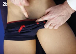
- Tenderness over pubic tubercle, superficial inguinal ring, conjoint tendon, distal rectus abdominis. Lifting the head helps locate landmarks
- Pain on resisted sit-up or Valsalva
- Palpate the superficial inguinal ring with the patient standing
Treatment
- Same general principles as adductor-related pain
- Abdominal and oblique strengthening, including eccentric work in the outer range
- Surgery for failed conservative care: laparoscopic or open posterior wall repair with mesh, sometimes combined with adductor tenotomy
4) Pubic-Related Groin Pain
Pubic bone stress at the symphysis. Formerly “osteitis pubis”. Symphyseal changes on imaging are common in asymptomatic athletes.
In athletes under about 20, consider pubic apophysitis.
Clinical features
- Central groin pain over the symphysis
- Often diffuse
- Localised tenderness of the symphysis and adjacent pubic bone
Imaging
- X-ray: “moth-eaten” appearance with erosions, sclerosis and osteophytes; check for symphyseal instability on flamingo views
- MRI: parasymphyseal bone marrow oedema
Treatment
- Same general principles as adductor-related pain; load management is key
- Refractory cases only: corticosteroid injection, dextrose prolotherapy, bisphosphonates, surgery
General Management
Acute injuries
- Initial protection, relative rest, ice, compression
- Early mobilisation and progressive rehabilitation
- Return to play: 2 weeks to 6 months, depending on structure and severity. Adductor injuries generally take longer than hip flexor injuries
Longstanding (overuse) injuries
- Majority of cases. Pain and disability usually progressive
- Targeted, progressive exercise therapy combined with cross training (upper body work, cycling)
- Early recognition and removal from the provoking load shortens time to return
- Analgesia to keep playing masks symptoms and tends to worsen the condition
- NSAIDs and corticosteroid injections are not routinely recommended
Prevention
- Copenhagen Adductor exercise (Adductor Strengthening Programme): reduces the prevalence of groin problems in male footballers by around 40%
- FIFA 11+ warm-up programme
- Adductor and hip flexor strengthening built into regular training
- Monitor training load, especially in pre-season
- Routine adductor squeeze strength screening can identify at-risk players
References
- Weir, A., Brukner, P., Delahunt, E., et al. (2015). Doha agreement meeting on terminology and definitions in groin pain in athletes. British Journal of Sports Medicine, 49(12), 768-774. doi:10.1136/bjsports-2015-094869
- Holmich, P., Uhrskou, P., Ulnits, L., et al. (1999). Effectiveness of active physical training as treatment for long-standing adductor-related groin pain in athletes: randomised trial. The Lancet, 353(9151), 439-443. doi:10.1016/S0140-6736(98)03340-6
- Haroy, J., Clarsen, B., Wiger, E. G., et al. (2019). The Adductor Strengthening Programme prevents groin problems among male football players: a cluster-randomised controlled trial. British Journal of Sports Medicine, 53(3), 150-157. doi:10.1136/bjsports-2017-098937
Related
Medical disclaimer: this article is general information, not personal medical advice. Always consult a qualified doctor about your own situation.