
Femoroacetabular Impingement
Definition
Abnormal, premature contact between the proximal femur and the acetabular rim during hip motion. May lead to labral tears and chondral damage.
FAI syndrome (Warwick Agreement 2016) is a clinical diagnosis requiring a triad:
- Symptoms: motion- or position-related hip/groin pain
- Signs: positive clinical tests, reduced range of motion
- Imaging: cam and/or pincer morphology
Morphology alone is not a diagnosis. Many people with cam or pincer shapes NEVER develop symptoms.
Epidemiology
- Cam morphology is common: roughly 1 in 4 to almost half of the general population on imaging, depending on definition used
- Higher in athletes from “hip-heavy” sports: football, ice hockey, basketball, ballet, martial arts, gymnastics
- FAI syndrome (symptoms + signs + imaging) is far less common, around 3% of adults aged 20 to 49
- Cam: young, athletic males
- Pincer: active, middle-aged females
- Mixed cam + pincer is the MOST COMMON pattern seen clinically
Relevant Anatomy
- Ball-and-socket joint: Femoral head should be spherical, with a concave head-neck junction that clears the rim in flexion
- Acetabular labrum: fibrocartilage ring on the acetabular rim. Deepens the socket, maintains a fluid seal, rich in nociceptors
- Chondrolabral junction: where labrum meets articular cartilage. The main site of damage in FAI
- Anterosuperior acetabulum (12 to 3 o’clock) is the usual zone of conflict, since this is what the femoral neck meets in flexion and internal rotation
Pathophysiology
- Repeated contact between femoral head-neck junction and acetabular rim during flexion and internal rotation
- Wolff’s law: bone remodels according to load. High-load hip activity during adolescence, while the proximal femoral physis is still open, is thought to drive cam formation
- Repetitive impingement leads to labral injury, then cartilage delamination, then secondary osteoarthritis
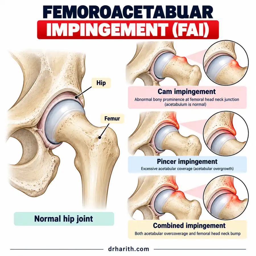
Classification
| Cam | Pincer | |
|---|---|---|
| Origin | Femoral | Acetabular |
| Typical patient | Young athletic male | Active middle-aged female |
| Morphology | Aspherical femoral head, bony bump at anterolateral head-neck junction | Acetabular over-coverage (focal retroversion or global, e.g. coxa profunda, protrusio) |
| Mechanism | Bump jams into the socket, shearing at the chondrolabral junction | Femoral neck abuts the overhanging rim, crushing the labrum |
| Damage pattern | Cartilage delamination, labral detachment (outside-in) | Intrasubstance labral tear, contrecoup posteroinferior cartilage lesion |
Mixed: features of both. Most common.
Clinical Presentation
History
- Insidious onset anterior hip or groin pain. Sometimes lateral hip or buttock
- C-sign: patient cups the hip between thumb and index finger
- Worse with activity, especially deep flexion and rotation
- Classic triggers: getting in and out of a car, prolonged sitting, squatting, putting on shoes and socks
- Mechanical symptoms: catching, clicking, locking (suggests labral tear)
- Stiffness and reduced performance in sport
Examination
- Reduced hip flexion (often less than 90 to 100°)
- Reduced internal rotation at 90° flexion. The most consistent sign
- FADIR test (flexion, adduction, internal rotation): reproduces groin pain. Highly sensitive, poorly specific. A negative test makes FAI syndrome unlikely
- FABER test: may reproduce groin or posterior pain, with reduced range compared to the other side
- Gait and hip muscle strength: abductor, extensor and rotator weakness is common
Investigations
Plain radiographs (first line)
- AP pelvis
- Cam: pistol grip deformity, loss of head-neck offset
- Pincer: crossover sign (acetabular retroversion), posterior wall sign, ischial spine sign
- Lateral centre-edge angle above about 40° suggests over-coverage
- Assess joint space for OA
- Lateral view: modified Dunn (45° flexion) or cross-table lateral. The view of choice for showing cam at the anterolateral head-neck junction
- Alpha angle: the most used cam measure. Commonly above 55 to 60° considered abnormal
CT
- 3D bony morphology, femoral version, surgical planning
MRI / MR arthrogram
- Labral tears, chondral damage, bone oedema
- MR arthrogram improves labral and cartilage detection
Diagnostic intra-articular injection
- Image-guided local anaesthetic. Pain relief confirms an intra-articular source
Associated Injuries
- Acetabular labral tear
- Chondral delamination
- Secondary hip osteoarthritis
- Compensatory overload of surrounding structures: adductors, hip flexors, pubic symphysis, lumbar spine
Differential Diagnosis
- Hip instability / isolated labral tear (e.g. dysplasia)
- Slipped upper femoral epiphysis (SUFE): adolescents, typically 12 to 15 years, overweight boys. Can present as knee pain. Always consider in a limping teenager
- Avascular necrosis of the femoral head
- Hip osteoarthritis
- Stress fracture of the femoral neck
- Sports-related groin pain (Doha classification): adductor-, iliopsoas-, inguinal- and pubic-related
- Referred pain: lumbar spine, sacroiliac joint
Management
No single proven algorithm. Shared decision-making based on age, symptoms, morphology, cartilage status and sporting goals.
Non-operative (first line)
- Education and activity modification: reduce provocative loading, especially sprinting, change of direction, kicking and deep flexion
- Physiotherapist-led, supervised, progressive exercise (e.g. Personalised Hip Therapy, 12 to 26 weeks)
- Hip abductor, extensor and rotator strengthening
- Start in isolated positions (often prone) for specificity, then progress to functional weight-bearing
- Core strengthening
- Balance and proprioception
- Modified training: stationary bike, pool-based work
- Analgesia: short-term NSAIDs
- Image-guided intra-articular injection: diagnostic, short-term therapeutic adjunct
Operative
Consider after failure of a structured programme of about 3 months, in patients without significant OA.
Prognosis
- Many respond to structured rehabilitation
- Cam morphology is a recognised risk factor for early hip osteoarthritis
- Symptomatic FAI syndrome with cam morphology carries a high 10-year risk of radiographic OA
- Return to sport after arthroscopy is generally high in athletes, but reduced if established cartilage damage
References
- Griffin, D. R., Dickenson, E. J., O'Donnell, J., et al. (2016). The Warwick Agreement on femoroacetabular impingement syndrome (FAI syndrome): an international consensus statement. British Journal of Sports Medicine, 50(19), 1169-1176. doi:10.1136/bjsports-2016-096743
- Griffin, D. R., Dickenson, E. J., Achana, F., et al. (2022). Arthroscopic hip surgery compared with personalised hip therapy in people over 16 years old with femoroacetabular impingement syndrome: UK FASHIoN RCT. Health Technology Assessment, 26(16). doi:10.3310/FXII0508
- Weir, A., Brukner, P., Delahunt, E., et al. (2015). Doha agreement meeting on terminology and definitions in groin pain in athletes. British Journal of Sports Medicine, 49(12), 768-774. doi:10.1136/bjsports-2015-094869
Related
Medical disclaimer: this article is general information, not personal medical advice. Always consult a qualified doctor about your own situation.